By Liz Chukwulebe
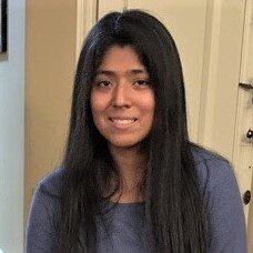
Melissa Lucero is a fifth-year PhD student studying chemistry and chemical biology. Lucero focuses on developing chemical tools to study disease states and biological processes via in vivo molecular imaging in the lab of chemistry professor Jefferson Chan, who is also a professor at the Beckman Institute for Advanced Science and Technology. The applications of Lucero's work range from studying aging to companion diagnostics and therapy. She is also the recipient of the Professor Gary Schuster Mentoring Scholarship in the Department of Chemistry.
Hometown: Silver Spring, MD
When did you first take an interest in your field?
My interest in molecular imaging began when I was an undergraduate researcher at the University of Maryland, Baltimore County. As an undergraduate, I learned how to synthesize complex molecules. However, I did not have the chance to apply the molecules to in vivo imaging. I learned that in vivo imaging was being used for fluorescence-guided surgery and therapies. Since I wanted to learn how to apply these chemical tools, I decided to join a research lab that performs biological research during a summer internship. This was an interesting experience for me because I saw how chemical tools, like fluorophores, could be used to study brain cells.
What kind of research are you working on?
My current work involves developing chemical tools to detect micrometastasis in the brain via photoacoustic imaging. Photoacoustic imaging is a hybrid of optical and ultrasound imaging that can provide high spatial resolution images in deep tissue. The way we are able to detect cancer is by designing our molecules to undergo a reaction with a specific analyte or biomarker that is elevated in cancer. Recently, this approach has enabled us to develop a prodrug-like molecule for lung cancer. In addition, I am studying changes in metal ion levels in the brain to see how they relate to aging.
How does your research make our world better?
The research I perform in the Chan Lab showcases the potential of photoacoustic imaging in a variety of settings, such as biological and pre-clinical areas. Ultimately, we believe that photoacoustic imaging is a safe, accessible, and relatively affordable mode of imaging that can be useful in the clinic for diagnosing and monitoring diseases.
How has your affiliation with the Beckman Institute helped you?
Much of the work I mentioned relied on the resources and equipment at the Beckman Institute. Standard murine models of cancer use subcutaneous (under the skin) tumors, which allows monitoring of tumor growth to be more accessible. However, this is an unnatural environment for most cancer cells, such as lung cancer. Therefore, I learned to implant cells in the pleural cavity of a mouse. To monitor the tumor burden via bioluminescence imaging, I frequently used the In Vivo Imaging System® SpectrumCT in the molecular imaging laboratory at the Beckman Institute.
Tell us about your post-university plans.
After graduating from the University of Illinois Urbana-Champaign, I will continue to work on molecular imaging with a focus on fluorescence as a postdoc at the National Cancer Institute in Frederick, MD. I believe that this position will allow me to gain more experience in using imaging modalities that have found use in the clinic.
What do you like to do outside of the classroom or lab?
When I am not focusing on lab work, I like to play and collect Rubik’s cubes which also help to alleviate some stress.
Favorite local restaurant: Sticky Rice
